Please get in contact with us if you are interested in any specific data sets or resources
Neonatal HRFs

The haemodynamic response in neonates, and in particular preterm neonates has markedly different morphology and temporal characteristics to the canonical adult function. The lag to positive peak time is significantly delayed and there is a deeper undershoot at term equivalent age. If you are analysing task based BOLD fMRI data, we strongly suggest that you consider using an age appropriate HRF model for convolution into your general linear model.
Please refer to these references if you are using this data or optimising you paradigm with it:
- Tomoki Arichi, Gianlorenzo Fagiolo, Marta Varela, Alejandro Melendez-Calderon, Alessandro Allievi, Nazakat Merchant, Nora Tusor, Serena J Counsell, Etienne Burdet, Christian F Beckmann, A David Edwards. Development of BOLD signal hemodynamic responses in the human brain Neuroimage 2012; 63(2): 663-73.
- Rhodri Cusack, Conor Wild, Annika C Linke, Tomoki Arichi, David SC Lee, Victor K Han. Optimizing stimulation and analysis protocols for neonatal fMRI. PloS One 2015; 10(8): e0120202.
Neonatal HRF as a function of age expand
by G.Fagiolo and T.Arichi
Data information expand
These are calculated from the data of 54 subjects between 32 and 44 weeks post-menstrual age using somatosensory stimulation of the right hand. Please quote the paper in reference 1.
Select age group between 32 and 38 weeks (press ENTER if necessary):
References expand
- Arichi, T., Fagiolo, G., Varela, M., Melendez-Calderon, A., Allievi, A., Merchant, N., Tusor, N., Counsell, S.J., Burdet, E., Beckmann, C.F., Edwards, A.D., 2012. Development of BOLD signal hemodynamic responses in the human brain. Neuroimage. available here
- RGraph for HTML5 plotting available here
Activation maps
Functional activation maps are available for download from http://brain-development.org/activation-maps/
- These include the preterm infant homunculus https://brain-development.org/somatotopicmap/ described in: Dall'Orso et al.
Somatotopic mapping of the developing sensorimotor cortex in the preterm human brain. Cerebral Cortex 2018; 28(7): 2507-15.

- Group activation maps of the fMRI clusters https://brain-development.org/activation-maps-related-to-spontaneous-delta-brush-activity/ associated with posterior-temporal spontaneous delta brush events in preterm infants described in Arichi et al. Localization of spontaneous bursting neuronal activity in the preterm human brain with simultaneous EEG-fMRI Elife 2017; 6: e27814.

The developing Human Connectome Project (dHCP)

Our group has also been involved in the ERC funded dHCP in collaboration with colleagues from King's College, Oxford University, and Imperial College London. A summary article of the neonatal data release including a description of the study population, image acquisition & processing, and collateral data (clinical and sociodemographic information, neurodevelopmental assessments, genetics) can be found in:
The work included a state-of-the-art resting state fMRI data processing pipeline led by Sean Fitzgibbon which was decribed in Fitzgibbon et al. Neuroimage 2020.
Resting state networks at term equivalent age from 337 infants in the dHCP have been described in Eyre et al. Brain 2021
The data for the dHCP including high resolution anatomical data, 300 direction multishell diffusion MRI data, high spatial and temporal resolution resting state fMRI data, and clinical & sociodemographic data is available for download.
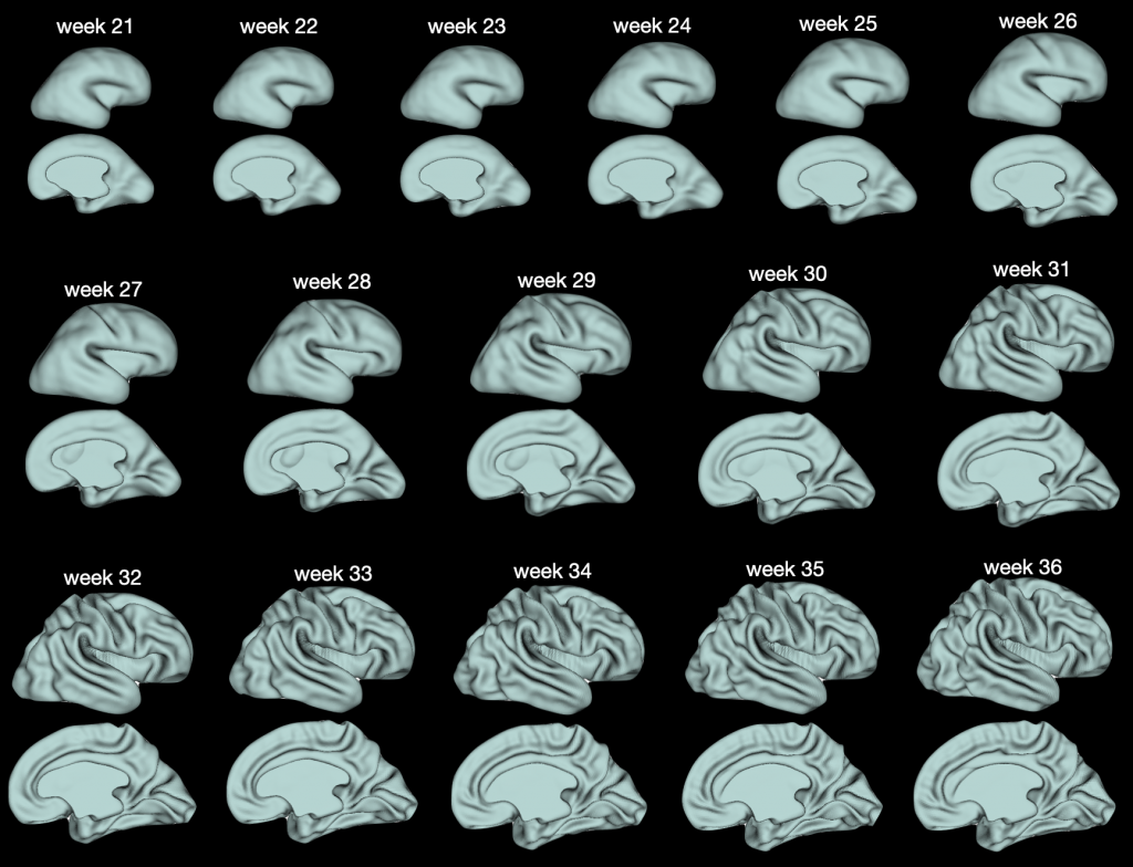
Fetal brain surface template
Fetal brain surface atlas, weeks 21-36 of gestation, constructed using 242 in-utero scans from the fetal dHCP cohort.
The following surfaces were generated: pial, mid-thickness, white matter and very inflated. The maps of changes in cortical morphology - sulcal depth, curvature, and thickness - are also available

An abstract describing this work has been submitted to OHBM 2023. Further details and an appropriate link to citation will follow. For any issues please write to: slava.karolis@kcl.ac.uk
Respect 4 Neurodevelopment Neurotechnology Network

Our group is involved in this inclusive multidisciplinary community that brings together neurodivergent families, bioengineers, psychologists, psychiatrists, paediatricians and related disciplines to develop Responsible, Reliable, Scalable and Personalised Neurotechnologies for neurodivergent children. More details about the network can be found here


